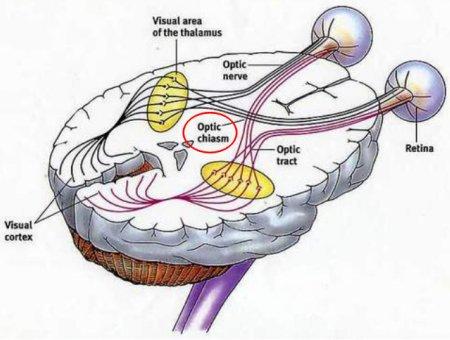
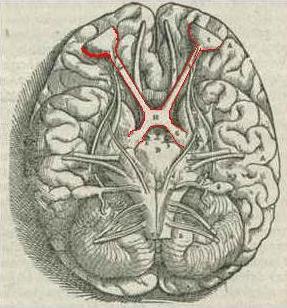
Anatomicalstructure
Opticalchiasm:Theopticchiasmbelongstothehypothalamusandislocatedontheundersideofthebrain.Thepartoftheopticchiasmthatstretchesforwardandoutwardiscalledtheopticnerve,whichconnectstotheeyeball;thefiberbundlethatstretchesouttothebackiscalledtheoptictract.Thatistosay,theopticchiasmiscomposedoffibersinsidetheskulloftheleftandrightopticnerves.Whenthefiberswithintheopticnervereachtheopticchiasma,thefiberintersectionofthenasal(medial)halfoftheleftandrightopticnervesoccurs,andthetemporal(lateral)halfThefibersofthepartdonotcross,andextendintotheoptictractaftercross.Therefore,oneopticbundlecontainsthefibersofthelateralhalfoftheipsilateralopticnerveandthemedialhalfofthecontralateralopticnerve.
Components
Thecomponentsoftheopticpathwaythatformtheoptictractaftertheopticnervefibercrossespartially:
Thenervefibersfromthetemporalhalfoftheretinadirectlyentertheipsilateraloptictractwithoutcrossing;thenervefibersfromthenasalhalfcrosstothecontralateraloptictract.Locatedbehindthetuberosityofsella,bloodissuppliedbythebranchesofthearterialring.Whenthemiddlepartoftheopticchiasmitselfisdamaged,itcausestemporalhemianopiaofbotheyes,whichismorecommoninthecompressionofpituitarytumorsandcraniopharyngiomas;lateraldamagecauseshemianopiaononesideofthenose,whichismorecommonininternalcarotidarteryatherosclerosis.


Themorphologicalstructureoftheopticchiasmanditsphysiologicalfunction
Theopticchiasmistheenlargedpartoftheopticnervechiasmjunctiononbothsides.Itislocatedabovethesellaandisellipticalorflatquadrilateral.Thethicknessisabout3to5mm,withanaverageof4mm,thefrontandreardiameteris4to13mm,withanaverageof8mm,andthetransversediameteris10to20mm,withanaverageofabout13.3mm.Thepositionoftheopticchiasmonthesaddleseptumvariesfrompersontoperson.Innormalhumans,5%arelocatedinthesphenoidnervegroove,12%areabovethesaddleseptum;theposterioredgeoftheopticchiasmisabovethesaddlebackaccountedfor79%,andthoselocatedbehindthesaddlebackaccountfor4%.Therelationshipbetweentheopticchiasmandsurroundingtissuesisveryimportant.Belowtheopticchiasm,thepituitaryglandisseparatedbythesaddleseptum.Thepituitaryglandconnectsthepituitaryglandwiththegraynodulesinthelowerthalamusthroughthesaddleseptum.Thedistancebetweentheopticchiasmandthesaddleseptumis5-10mm,andthereistheopticchiasmcistern.Tumorsinthesaddlecannotdirectlycompresstheopticchiasmbeforebreakingthroughthesaddleseptum.Bothposteriorandinferiorareadjacenttotheopticcryptandfunnelcryptofthethirdventricle.Therefore,anyenlargementofthethirdventriclecausedbyanyreasoncancompresstheopticchiasmandcausevisualfielddefect;Connecthere.Thepositionoftheopticchiasmaisanterior,andtheanteriorcommunicatingarteryislocatedaboveit.Ifananeurysmoccursinthisartery,theopticchiasmiscompressed,andvisualfielddefectsintheinferiortemporalquadrantoftheeyesappear;behindtheopticchiasmisaquadrangularinterpodalcistern,bothsidesofthecerebralpeduncleandpituitaryglandThefunnelextendsalongtheposterioredgeoftheopticchiasmthroughthesaddleseptumandextendsintothesaddle,andiscontinuouswiththeposteriorpituitary;thesideoftheopticchiasmisadjacenttotheinternalcarotidarteryandposteriorcommunicatingartery,andthecavernoussinusisoutsideandbelowit.Therefore,inadditiontodiseasesoftheopticchiasmitself,diseasesinitssurroundingareascanofteninvadetheopticchiasmandcausecorrespondingvisualfielddefects,whichcanbeusedasabasisfordiagnosis.Thenervefibersintheopticchiasmincludetwogroups,crossedandnon-crossed.Thecrossedfiberscomefromthenasalhalfoftheretinaofbotheyes;thenon-crossedfiberscomefromthetemporalhalfoftheretinaofbotheyes.Thecrossfibersfromtheupperhalfoftheretinaoccupytheupperlayeroftheopticchiasm,formingtheposteriorkneeonthesameside,andthengototheoppositeopticbundle;thecrossfibersfromthelowerhalfoccupythelowerlayeroftheopticchiasm,formingtheanteriorkneeontheoppositeside,andentertheoppositesideOpticbeam.Thenon-crossingfibersfromtheupperhalfoftheretinaarelocatedinsideandabovethesamesideoftheopticchiasm,andthenon-crossingfibersfromthelowerhalfarelocatedonthesamesideandbelowandoutsidetoentertheipsilateraloptictract.Macularfibersarealsodividedintotwotypes:crossedandnon-crossed.Thecrossedmacularfiberscrosstotheoppositesideoftheopticchiasm;thenon-crossedmacularfibersenterthesamesideoftheopticbundle.Therefore,differentvisualfieldchangescanoccurindifferentpartsoftheopticchiasm,whichisofgreatclinicalsignificance.
Diseasesrelatedtotheopticchiasm
1.Opticchiasmglioma:
OpticalgliomaisoneofthemostcommonprimarytumorsintheopticpathwayAccordingtotheanatomicallocation,itcanbedividedintointraorbitaltype,cranioorbitalcommunicationtypeandintracranialtype.Theintracranialtypecanbedividedintothreetypes:typeⅠ,pureopticnervetype;typeⅡ,pureopticchiasmortypeinvolvingtheopticnerveatthesametime;typeⅢ,involvinghypothalamusorotherstructuraltypes.Themainclinicalsymptomsarevisionloss,visualfielddefectandopticnerveatrophy.Ifthehypothalamusisinvolved,theremaybeobesity,growthretardationandprecociouspuberty,sexualdysfunctionandmenstrualdisorders.
Intracranialopticnerve-opticchiasmgliomahasmorecharacteristicimagingfindings.MRIshowsthatthelesionissignificantlybetterthanCT.OnplainCTscan,thelesionsareiso-densityorslightlylow-density,andsomeofthemgrowalongthesideoftheopticnerve.Itisdifficulttoshowthenormalopticchiasmstructure.EnhancedCTscansusuallyshowuniformandmoderateenhancement.MRImainlymanifestsaslongT1andlongT2signallesionsonthesaddle,withclearborders,andtheopticnerveor(and)theopticchiasmisthickenedorenlargedinafusiformormassshape,andasmallpartofitmayhavecysticdegenerationandcalcification.EnhancedscanningItismoderatelyuniformlystrengthened.
2.Opticchiasminjury:damagetotheforeheadismostlycausedbyforce,andcompressionofbonefragmentsisrare.Vasculardamagemaybeoneoftheimportantreasons.Differentvisualfielddefectsandfunduschangescanoccurduetodifferentinjurysitesandscopes.
Tumorsaroundtheopticchiasm
Intracranialcongenitaltumors:
1.Epidermoidcystsanddermoidcysts:
(一)Histogenesis
Epidermoidcystsanddermoidcystsaregenerallyconsideredtodevelopfromtheappendagesoftheskinectoderm.Thisattachmentisembeddedinthebrainwhentheneuraltubebreaksawayfromtheectodermandclosesintheearly3to5weeksofembryonicdevelopment,andectopicresidueoccurs.Iftheectopicoccursintheveryearlystage,whentheskinectodermhasnotyetdifferentiatedintovariousskinstructures,itwilldifferentiateintovariouscomponentsoftheskinafterbeingembeddedtoformdermoidcysts;forexample,theskinectodermcellshavedifferentiatedAfterbeingembedded,itonlydevelopsintoepidermaltissueandformsepidermoidcyst.Itisalsobelievedthatepidermoidcystsanddermoidcystshavenothingtodowiththedevelopmenttime.Ifalllayersoftheskinareectopic,itwilldevelopintoadermoidcyst;ifonlytheepidermaltissueisectopic,itwilldevelopintoanepidermoidcyst.Epidermoidcystcontainsonlyonegermlayer,whichistheectodermcomponent.Dermoidcystcontainstwogermlayers:ectodermandmesoderm.Teratomacontainsthreegermlayers.
2.Chordoma:Thetumoratthebaseoftheskullinitiallygrowsontheepidural,coveredwithacapsule,andthebottomisinfiltratedtodestroytheboneatthebaseoftheskullandinvadenerves.Sacrococcygealtumorsinfiltrateanddestroythevertebraeandintervertebralvertebrae.Thetumorsaremostlylobulated,withasmoothsurface,alubricitytothetouch,gray-white,varyinginquality,withclearboundariesintheearlystagesandunclearboundariesinthelatestages.Thecutsurfaceshowscystsofvaryingsizes,containingtranslucentjelly-likeormucous-likesubstances,andthefibroustissueisdividedintolobules,whichmaybecalcified,andoldbleeding,necrosis,andcysticdegenerationarecommon.Thosewithsofttumortissueproducemoremucusandtendtobebenign,whilethosewithhardtissuemayhavemorecalcificationandhaveagreatertendencytomalignancy.
